top of page

Gorman Images
For use in presentations only.
ABME does not own these images and permission to publish must be obtained from David Gorman.

External Oblique

Transversus Abdominis

Internal Oblique

Serratus Posterior Superior

Pectoralis Minor and Serratus Anterior)

Posterior view of the tongue in cross-section

Pterygoids

Progressive movement of mandibular condyle in the TMJ in opening and closing the mouth

digastrics and Sternothyroid, sternohyoid, omohyoid

Temporalis muscle with zygomatic arch cut to show insertion into coronoid process

Levator and Tensor veli palatini muscles

Lateral view of pharyngeal constrictors showing insertions into vocal tract structures)

Posterior view of pharynx with muscles cut on one side to show cavities in front)

Neck muscles, chin level, not lifted

Larynx lateral view, uncut)

Larynx lateral view, cut)

Glottal closure
Adduction

Abduction

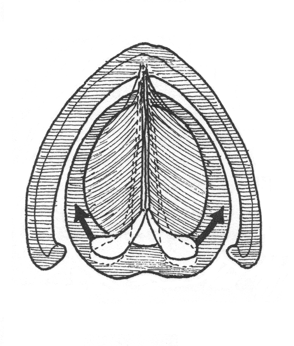
Vocal Ligaments Superior view)

Larynx posterior view

Mandible, cervical spine, hyoid, larynx trachea, clavicle, first rib

Ankle Joint Front View
bottom of page